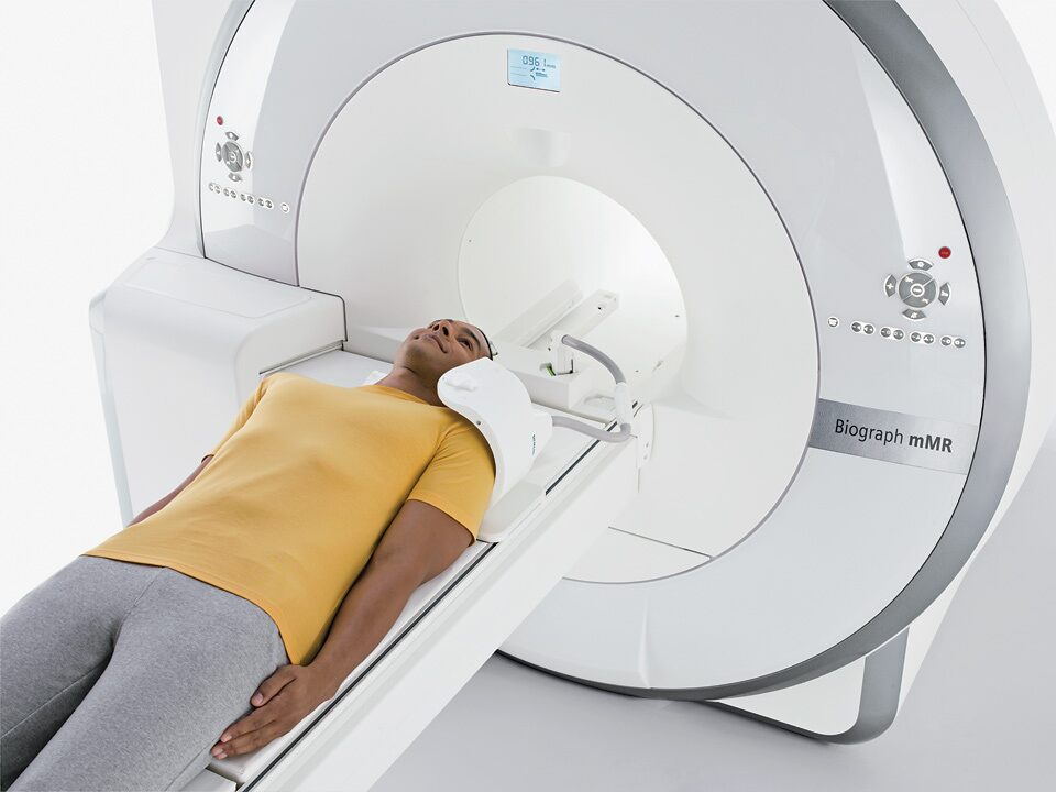
The Biograph mMR scanner (Siemens Healthineers, Erlangen, Germany) consists of a 3T whole-body superconductive magnet with active shielding and external interference shielding and a whole-body PET scanner. It is equipped with a gradient system with a maximum gradient amplitude of 45 mT/m and a maximal slew rate of 200 T/m/s. Separate cooling channels that simultaneously cool primary and secondary coils allow the application of extremely gradient intensive techniques.
This scanner is equipped with the “TIM” RF coils that were custom designed to minimize the 511 keV photons attenuation. The fully-integrated PET detectors use avalanche photodiode (APD) technology and LSO scintillator crystals (eight rings with 56 detectors blocks per ring, each consisting of 8×8 arrays of 4×4×20 mm3crystals read out by a 3×3 array of APDs). The PET scanner’s transaxial and axial fields of view are 594 mm and 25.8 cm, respectively.

The Biograph mMR was installed at the Martinos Center in June 2011.