Development of the Human Dynamic NeuroChemical Connectome (HDNCC) Scanner

Development of the Human Dynamic NeuroChemical Connectome (HDNCC) Scanner

The goal of this project is to design and build a 7-T MR-compatible PET camera with >10x improved sensitivity to enable dynamic PET imaging of brain neurotransmission, neuromodulation, and other dynamic molecular events with unprecedented temporal resolution and beyond state-of-the-art spatial resolution. This will allow us to merge the dynamic functional capabilities of both PET and MRI methods, providing investigators the unique capacity to perform experiments linking structure with electrical (through its surrogate hemodynamics) and neurochemical function on time scales relevant for understanding human cognition. 

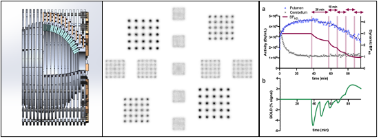
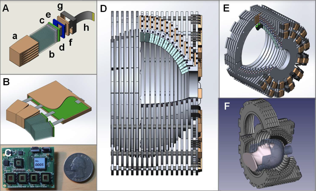
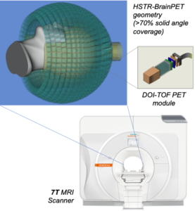
We will address the hardware and software challenges in assembling 7-T MR-compatible PET technology purpose-built to extend the temporal window of brain PET imaging down to just a few seconds. Funding for demonstrating proof-of-concept (i.e., develop the PET detectors and build a partial scanner) was provided by the BRAIN Initiative NIH-NIBIB&NINDS (1R01-EB026995-01; PI: Catana). We proposed to address the two main factors that determine PET sensitivity: geometric efficiency (to maximize the probability of photons to reach the detectors)and detection efficiency (to detect most of the incident photons).

Specifically, we will use a non-conventional spherical geometry to increase the solid angle coverage to ~71%. This change will translate into ~25% sensitivity for detecting true coincidences. Additionally, to decode the scintillator blocks we will design high-performance readout electronics with depth-of-interaction and time-of-flight (TOF, to improve the count rate performance) capabilities. Furthermore, the TOF information will also act as a virtual sensitivity amplifier and thus sensitivity could be as high as 50%, a dramatic improvement compared to current values (i.e. 1-2%).  

Our preliminary results to date suggest that:

  • very high sensitivity will indeed be obtained using the proposed partial-sphere PET geometry;
  • the photon detectors and associated electronics show no mutual interference with the 7-T system;
  • the 7-T main magnetic field will not be significantly perturbed by the PET scintillator arrays;
  • a high-performance transmit-receive 7-T MR array can be integrated into the PET gantry.

The recently awarded BRAIN Initiative grant U01EB029826 (PI: Catana) will provide funding to:

  1. Build the HSTR-BrainPET using 7-T MR-compatible technology to enable interference-free simultaneous data acquisition.
  2. Implement PET data acquisition and image reconstruction software for the spherical geometry.
  3. Apply the integrated HSTR-BrainPET & 7-T MRI scanner to the dynamic assessment of neurochemical events and brain activation in healthy subjects.

For the hardware/software developments proposed in this project, we have expanded our longstanding partnership with Siemens Healthineers by including experts from University of Tubingen (Germany), Hamamatsu (Japan), Complutense University of Madrid (Spain), and University of Texas at Arlington.

Siemens BrainPET

The BrainPET prototype (Siemens Healthineers) is a head insert designed to fit inside the 3T Siemens TIM Trio 60 cm whole-body MRI scanner. There are 32 detector cassettes that make up the BrainPET gantry, each consisting of six detector blocks. Each detector block consists of a 12×12 array of lutetium oxyorthosilicate (LSO) crystals (2.5×2.5×20 mm3) readout by a 3×3 array of Hamamatsu avalanche photodiodes (APDs, 5×5 mm2).  The gantry physical inner and outer diameters are 35 and 60 cm, respectively. The transaxial and axial fields-of-view are 32 cm and 19.125 cm, respectively.

After the BrainPET was installed at the Martinos Center in May 2008, we worked very closely with the Siemens engineers to optimize its performance and improve the image quality. We have been using the BrainPET in a myriad of studies ranging from those aimed at investigating the mutual interference between the two devices and the performance of the PET camera, developing methods to use the information obtained from one device to improve the other modality, and performing proof-of-principle studies in small animal, non-human primates and humans.

Relevant publications

Siemens Biograph mMR

The Biograph mMR scanner (Siemens Healthineers, Erlangen, Germany) consists of a 3T whole-body superconductive magnet with active shielding and external interference shielding and a whole-body PET scanner. It is equipped with a gradient system with a maximum gradient amplitude of 45 mT/m and a maximal slew rate of 200 T/m/s.  Separate cooling channels that simultaneously cool primary and secondary coils allow the application of extremely gradient intensive techniques. 

This scanner is equipped with the “TIM” RF coils that were custom designed to minimize the 511 keV photons attenuation. The fully-integrated PET detectors use avalanche photodiode (APD) technology and LSO scintillator crystals (eight rings with 56 detectors blocks per ring, each consisting of 8×8 arrays of 4×4×20 mm3crystals read out by a 3×3 array of APDs).  The PET scanner’s transaxial and axial fields of view are 594 mm and 25.8 cm, respectively.

https://www.siemens-healthineers.com/en-us/magnetic-resonance-imaging/mr-pet-scanner/biograph-mmr

The Biograph mMR was installed at the Martinos Center in June 2011.