Hardware

our research

HARDWARE

Develop, optimize and validate integrated PET/MRI hardware.

BRAIN
WHOLE BODY
ADD-ON DEVICES

brain

PET provides unique information about the human brain that has been proved useful in a variety of studies ranging from basic neuroscience to clinical applications. In parallel with the development of dozens of radiotracers and the implementation of methods for extracting ever more accurate estimates of parameters related to many biological processes, PET instrumentation has continuously evolved over the last six decades. In addition to general-purpose whole-body devices, several scanners designed specifically for studying the brain have also been developed, motivated by the need for improved performance, specifically better spatial resolution and/or higher sensitivity to enable the imaging of small brain structures. Furthermore, given the smaller overall size, the integration with MRI scanners is also easier and this and other non-technical reasons have led to the development of MR-compatible PET inserts. The latest developments in dedicated brain PET imaging hardware were reviewed in [Catana 2019]. 

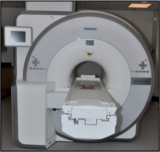
The BrainPET prototype (Siemens Healthineers) is a head insert designed to fit inside the 3T Siemens TIM Trio 60 cm whole-body MRI scanner. There are 32 detector cassettes that make up the BrainPET gantry, each consisting of six detector blocks. Each detector block consists of a 12×12 array of lutetium oxyorthosilicate (LSO) crystals (2.5×2.5×20 mm3) readout by a 3×3 array of Hamamatsu avalanche photodiodes (APDs, 5×5 mm2).  The gantry physical inner and outer diameters are 35 and 60 cm, respectively. The transaxial and axial fields-of-view are 32 cm and 19.125 cm, respectively.

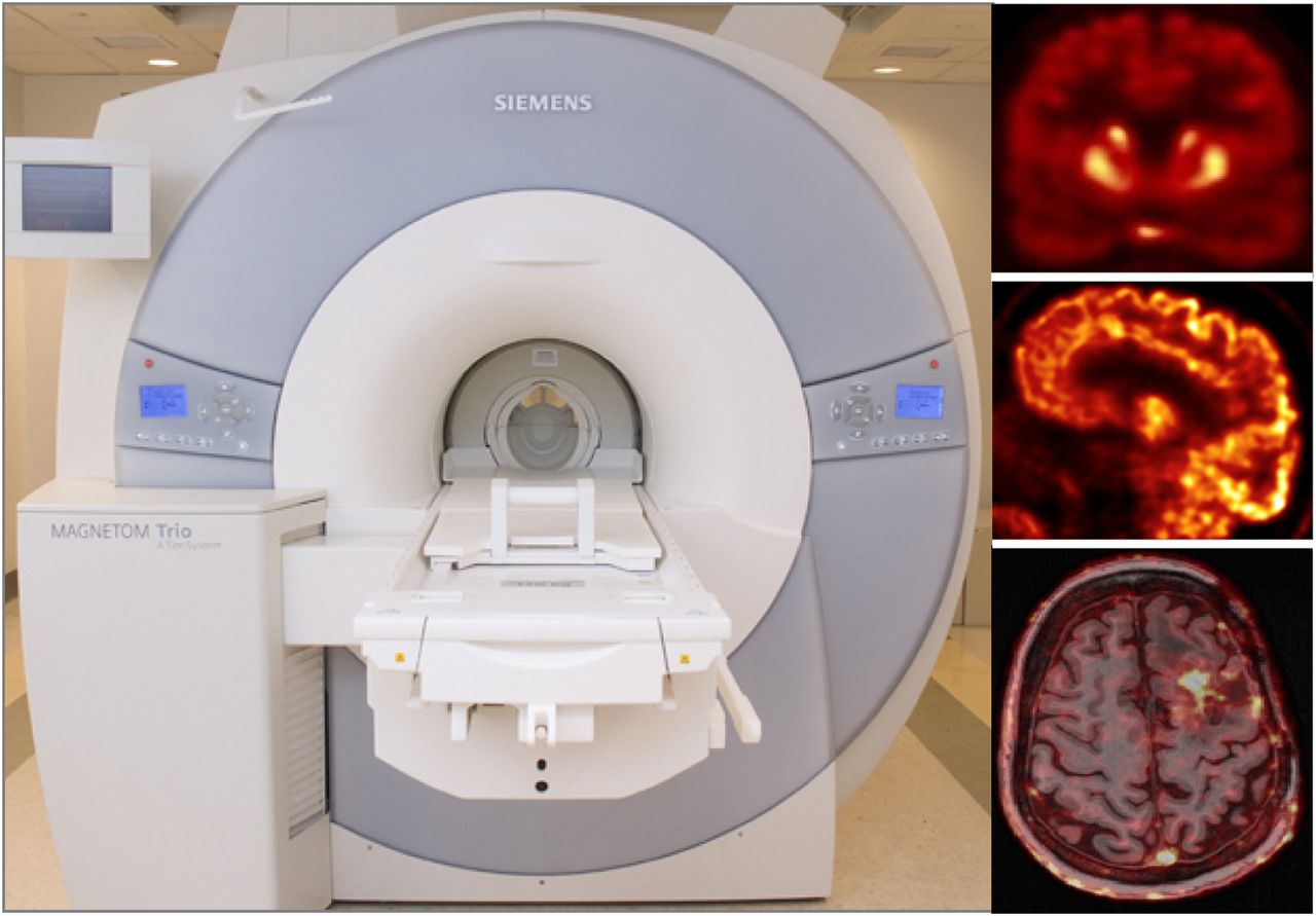
After the BrainPET was installed at the Martinos Center in May 2008, we worked very closely with the Siemens engineers to optimize its performance and improve the image quality. We have been using the BrainPET in numerous studies ranging from those aimed at investigating the mutual interference between the two devices and the performance of the PET camera, developing methods to use the information obtained from one device to improve the other modality, and performing proof-of-principle studies in small animal, non-human primates and humans.

We are currently leading an effort with our collaborators from Siemens and several other institutions around the world to build the next generation integrated system with an order of magnitude improvement in sensitivity.

Whole
Body

We were the first US adopters of the Biograph mMR scanner (Siemens Healthineers, Erlangen, Germany). This scanner consists of a 3T whole-body superconductive magnet with active shielding and external interference shielding and a whole-body PET scanner. It is equipped with a gradient system with a maximum gradient amplitude of 45 mT/m and a maximal slew rate of 200 T/m/s. Separate cooling channels that simultaneously cool primary and secondary coils allow the application of extremely gradient intensive techniques. This scanner is equipped with the “TIM” RF coils that were custom designed to minimize the 511 keV photons attenuation. The fully-integrated PET detectors use avalanche photodiode (APD) technology and LSO scintillator crystals (eight rings with 56 detectors blocks per ring, each consisting of 8×8 arrays of 4×4×20 mm3 crystals read out by a 3×3 array of APDs). The PET scanner’s transaxial and axial fields of view are 594 mm and 25.8 cm, respectively. 


Siemens Biograph mMR scanner installed at the Martinos Center in 2011

Add-on
devices

Arterial sampling by hand is time consuming, prone to errors, and unnecessarily subjects study staff to additional radiation. As an alternative an MR-compatible automated blood sampler was built by Dan Chonde following the work of Breuer et al.