Motion correction
MR-based for brain studies
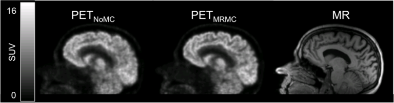
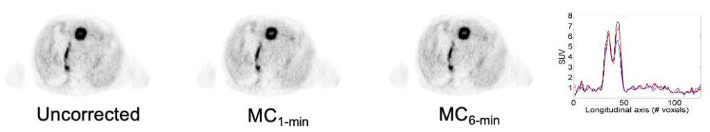
Head motion is difficult to avoid in long PET studies, degrading the image quality and offsetting the benefit of using a high-resolution scanner. As a potential solution in an integrated MR-PET scanner, the simultaneously acquired high-temporal resolution MR data can be used for motion tracking. We proposed a data processing and rigid-body motion correction algorithm for the MR-compatible BrainPET prototype scanner and performed proof-of-principle phantom and human studies (Catana 2011).
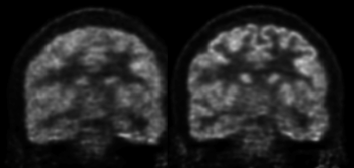
This method was subsequently used in research studies aimed at assessing the role of dopamine D1 signaling in working memory (Roffman 2016) and the role of central dopamine in human bonding (Atzil 2017). More recently, we showed the variability in the PET estimation of the cerebral metabolic rate of glucose utilization is reduced after MR-assisted motion correction in Alzheimer’s disease patients (Chen 2018).
PET-based for brain studies
When MR-derived motion estimates are unavailable, either in the gaps between sequences or when a given MR sequence is not amenable to motion estimation, motion can also be derived directly from the PET images themselves. Motion estimates from both the MR and the PET can be unified to provide continuous estimates of head motion throughout the duration of the scan, leveraging the higher resolution and more accurate MR-based estimates when they are available. (Levine 2017 Abstract)
MR-based for cardiac studies
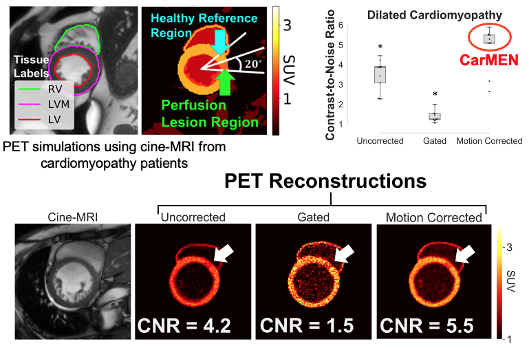
We proposed an unsupervised deep learning-based approach for deformable three-dimensional cardiac MR image registration. This method learns a motion model that balances image similarity and motion estimation accuracy. We validated our approach comprehensively on three datasets and demonstrated higher motion estimation and registration accuracy relative to several popular state-of-the-art image registration methods (Morales 2019).
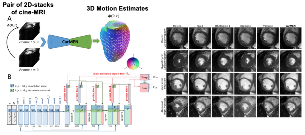
When used for PET motion correction, CarMEN led to an increase in the contrast-to-noise ratio in the simulated perfusion lesions (Morales et al, ISMRM/SNMMI co-provided PET/MRI Workshop, New York 2019).
MR-based for abdominal studies
PET and MRI are two powerful imaging technologies that are characterized by high sensitivity and the ability to provide superior anatomic detail, respectively, which might make them ideal for evaluating the upper abdomen. However, PET requires long acquisition times, including the acquisition of data from moving organs, which may result in image blurring. On the other hand, MRI, especially standard DCE-MRI, can scan the chosen field of view in a shorter time, but requires the patient’s cooperation with the respiratory instructions and the ability to suspend respiration for the acquisition time of breath-hold sequences, usually in the range 14–20 s. Moreover, even in patients with an adequate respiratory breath-hold ability, the quality and the diagnostic information of DCE-MRI are also dependent on the hemodynamics of the patient and the timing of contrast agent injection and data acquisition. These variables explain the occurrence of respiratory artifacts and erroneous phases of contrast enhancement imaging in DCE-MRI.
We presented and evaluated in vivo a comprehensive approach for self-gated MR motion modeling applied to concurrent respiratory motion compensation of PET and DCE-MRI data acquired simultaneously in an integrated PET/MR system.
Fully registered, motion-corrected PET images and diagnostic DCE-MR images were obtained with negligible acquisition time prolongation compared with standard breath-hold techniques. Both the MR and the PET image quality and tracer uptake quantification were improved when compared with conventional methods (Fuin 2018).
This approach was subsequently evaluated clinically in collaboration with Dr. Onofrio Catalano to demonstrate that motion-corrected PET/MRI produced better PET images and reduced the spatial mismatch between the two modalities (Catalano 2018).