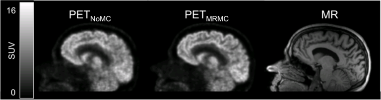
Head motion is difficult to avoid in long PET studies, degrading the image quality and offsetting the benefit of using a high-resolution scanner. As a potential solution in an integrated MR-PET scanner, the simultaneously acquired high-temporal resolution MR data can be used for motion tracking. We proposed a data processing and rigid-body motion correction algorithm for the MR-compatible BrainPET prototype scanner and performed proof-of-principle phantom and human studies(Catana 2011).
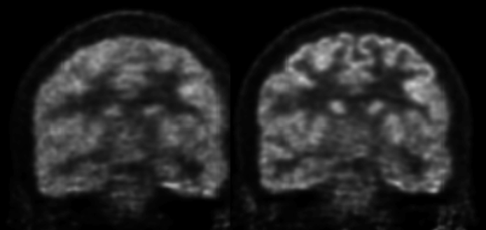
FDG-BrainPET images before (left) and after (right) MR-assisted motion correction
This method was subsequently used in research studies aimed at assessing the role of dopamine D1 signaling in working memory (Roffman 2016) and the role of central dopamine in human bonding (Atzil 2017). More recently, we showed the variability in the PET estimation of the cerebral metabolic rate of glucose utilization is reduced after MR-assisted motion correction in Alzheimer’s disease patients (Chen 2018).