Attenuation correction for pelvis PET imaging

Attenuation correction for pelvis PET-MR imaging

The pelvis is another area where MR-based approaches that do not properly account for bone attenuation could lead to substantial bias in PET data quantification and deep learning approaches have been proposed to minimize this bias starting from the MR data acquired using specialized MR sequences. Instead, we implemented a deep learning-based approach to generate pseudo-CT maps exclusively from the Dixon-VIBE MR images routinely acquired for attenuation correction on the Biograph mMR scanner. Using our 2D network that takes four contrasts as inputs (water, fat, in-phase, and out-of-phase Dixon-VIBE images), the mean absolute relative change in PET values in the pelvis area was 2.36% ± 3.15% (Torrado-Carvajal et al 2019).

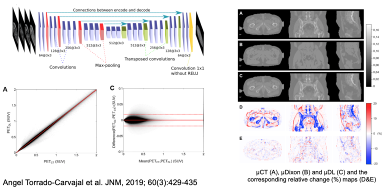
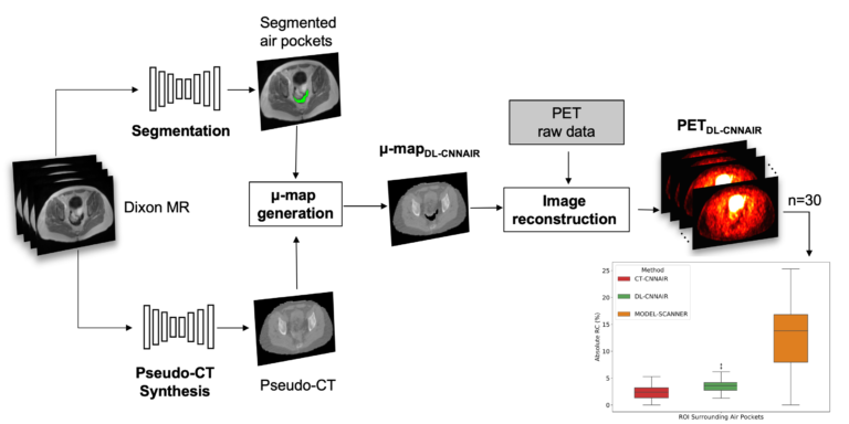
One remaining challenge in using convolutional neuronal networks to synthesize CT from MR images of the pelvis is the presence of air pockets (i.e., digestive tract gas) in this area. As CT and MR images are acquired on separate scanners at different times, the locations and sizes of these air pockets can change between the two scans, which can lead to errors in both the MR-CT co-registration and image synthesis tasks. We trained and evaluated CNNs to automatically segment air pockets from MR CAIPIRINHA-accelerated Dixon images and assessed the quantitative impact on the reconstructed PET images (Sari et al, submitted to JNM).

Attenuation correction for brain PET imaging

Attenuation correction for brain PET-MR imaging studies 

Identifying bone tissue is particularly relevant for accurate attenuation correction in neurological PET studies as this tissue class has the highest linear attenuation coefficient and inaccuracies in its estimation can introduce large biases in the adjacently located cortical structures. Since using conventional MRI pulse sequences bone tissue and air-filled cavities are difficult to distinguish, novel sequences have been developed to address this challenge. We were among the first to propose using ultra-short echo time MR sequences (optimized for imaging tissues with very short T2 relaxation times) for generating segmented head attenuation maps.  In a first approach, the head was segmented into three compartments (i.e. bone and soft tissue and air cavities) based on the relationship between the two echoes on a voxel-by-voxel basis (Catana 2010). 

Subsequently, combining the dual-echo UTE and T1-weighted MR data using probabilistic atlases allowed the generation of substantially improved segmented attenuation maps  (Poynton, Chen et al 2014). However, one of the main limitations of early generation UTE-based methods was that only three compartments (i.e. soft tissue, bone, and air cavities) could be identified. 

Segmented head attenuation maps derived from CT (upper row) and T1w&DUTE MR data (lower row) (Poyton, Chen et al. 2014)

We next implemented an SPM-based method for generating head attenuation maps from a single morphological MRI dataset obtained with the MPRAGE sequence routinely collected in research neurological studies (Izquierdo-Garcia et al 2014). After intensity normalization, the MR images are segmented into six tissue classes using the “New Segment” SPM tool and then registered to a previously created template using a diffeomorphic non-rigid image registration algorithm (SPM DARTEL). The inverse transformation is applied to obtain the pseudo-CT images in the subject space. Using this approach, the voxel- and regional-based quantification errors compared to the scaled CT method were 3.87±5.0% and 2.74±2.28%, respectively. This method was also demonstrated to work in patients with modified anatomy (e.g. glioblastoma patients post-surgery). This method and several of the others developed by other groups around the world have been shown in a multi-center evaluation to be quantitatively accurate to a degree “smaller than the quantification reproducibility in PET imaging” (Ladefoged 2017). Continuous-valued attenuation maps were also obtained from dual-echo UTE and T1-weighted MR data using probabilistic atlases (Chen et al 2017).  We also assessed the repeatability of the atlas- or UTE-based methods.  For example, comparable attenuation maps and PET volumes were obtained at three visits using the probabilistic atlas method (Chen et al 2017).  Similar results were reported for the SPM-based method (Izquierdo-Garcia et al 2018).

Software

  • Masamune (includes several methods for generating head attenuation maps from MR or PET data)
  • Stand-alone PseudoCT for the generating head attenuation maps for BrainPET and Biograph mMR studies

Development of the Human Dynamic NeuroChemical Connectome (HDNCC) Scanner

Development of the Human Dynamic NeuroChemical Connectome (HDNCC) Scanner

The goal of this project is to design and build a 7-T MR-compatible PET camera with >10x improved sensitivity to enable dynamic PET imaging of brain neurotransmission, neuromodulation, and other dynamic molecular events with unprecedented temporal resolution and beyond state-of-the-art spatial resolution. This will allow us to merge the dynamic functional capabilities of both PET and MRI methods, providing investigators the unique capacity to perform experiments linking structure with electrical (through its surrogate hemodynamics) and neurochemical function on time scales relevant for understanding human cognition. 

We will address the hardware and software challenges in assembling 7-T MR-compatible PET technology purpose-built to extend the temporal window of brain PET imaging down to just a few seconds. Funding for demonstrating proof-of-concept (i.e., develop the PET detectors and build a partial scanner) was provided by the BRAIN Initiative NIH-NIBIB&NINDS (1R01-EB026995-01; PI: Catana). We proposed to address the two main factors that determine PET sensitivity: geometric efficiency (to maximize the probability of photons to reach the detectors)and detection efficiency (to detect most of the incident photons).

Specifically, we will use a non-conventional spherical geometry to increase the solid angle coverage to ~71%. This change will translate into ~25% sensitivity for detecting true coincidences. Additionally, to decode the scintillator blocks we will design high-performance readout electronics with depth-of-interaction and time-of-flight (TOF, to improve the count rate performance) capabilities. Furthermore, the TOF information will also act as a virtual sensitivity amplifier and thus sensitivity could be as high as 50%, a dramatic improvement compared to current values (i.e. 1-2%).  

Our preliminary results to date suggest that:

  • very high sensitivity will indeed be obtained using the proposed partial-sphere PET geometry;
  • the photon detectors and associated electronics show no mutual interference with the 7-T system;
  • the 7-T main magnetic field will not be significantly perturbed by the PET scintillator arrays;
  • a high-performance transmit-receive 7-T MR array can be integrated into the PET gantry.

The recently awarded BRAIN Initiative grant U01EB029826 (PI: Catana) will provide funding to:

  1. Build the HSTR-BrainPET using 7-T MR-compatible technology to enable interference-free simultaneous data acquisition.
  2. Implement PET data acquisition and image reconstruction software for the spherical geometry.
  3. Apply the integrated HSTR-BrainPET & 7-T MRI scanner to the dynamic assessment of neurochemical events and brain activation in healthy subjects.

For the hardware/software developments proposed in this project, we have expanded our longstanding partnership with Siemens Healthineers by including experts from University of Tubingen (Germany), Hamamatsu (Japan), Complutense University of Madrid (Spain), and University of Texas at Arlington.

Siemens BrainPET

The BrainPET prototype (Siemens Healthineers) is a head insert designed to fit inside the 3T Siemens TIM Trio 60 cm whole-body MRI scanner. There are 32 detector cassettes that make up the BrainPET gantry, each consisting of six detector blocks. Each detector block consists of a 12×12 array of lutetium oxyorthosilicate (LSO) crystals (2.5×2.5×20 mm3) readout by a 3×3 array of Hamamatsu avalanche photodiodes (APDs, 5×5 mm2).  The gantry physical inner and outer diameters are 35 and 60 cm, respectively. The transaxial and axial fields-of-view are 32 cm and 19.125 cm, respectively.

After the BrainPET was installed at the Martinos Center in May 2008, we worked very closely with the Siemens engineers to optimize its performance and improve the image quality. We have been using the BrainPET in a myriad of studies ranging from those aimed at investigating the mutual interference between the two devices and the performance of the PET camera, developing methods to use the information obtained from one device to improve the other modality, and performing proof-of-principle studies in small animal, non-human primates and humans.

Relevant publications

Siemens Biograph mMR

The Biograph mMR scanner (Siemens Healthineers, Erlangen, Germany) consists of a 3T whole-body superconductive magnet with active shielding and external interference shielding and a whole-body PET scanner. It is equipped with a gradient system with a maximum gradient amplitude of 45 mT/m and a maximal slew rate of 200 T/m/s.  Separate cooling channels that simultaneously cool primary and secondary coils allow the application of extremely gradient intensive techniques. 

This scanner is equipped with the “TIM” RF coils that were custom designed to minimize the 511 keV photons attenuation. The fully-integrated PET detectors use avalanche photodiode (APD) technology and LSO scintillator crystals (eight rings with 56 detectors blocks per ring, each consisting of 8×8 arrays of 4×4×20 mm3crystals read out by a 3×3 array of APDs).  The PET scanner’s transaxial and axial fields of view are 594 mm and 25.8 cm, respectively.

https://www.siemens-healthineers.com/en-us/magnetic-resonance-imaging/mr-pet-scanner/biograph-mmr

The Biograph mMR was installed at the Martinos Center in June 2011.